
recently, This lecture focuses on "artificial intelligence assists precise diagnosis and treatment of tumors". Focusing on the cutting-edge application of AI in the two key fields of tumor pathology and medical imaging, Professor Yang Baocheng of Wenzhou Medical University and Professor Xing Xiaohan of the National University of Singapore are specially invited to give special lectures. The academic atmosphere is strong 。
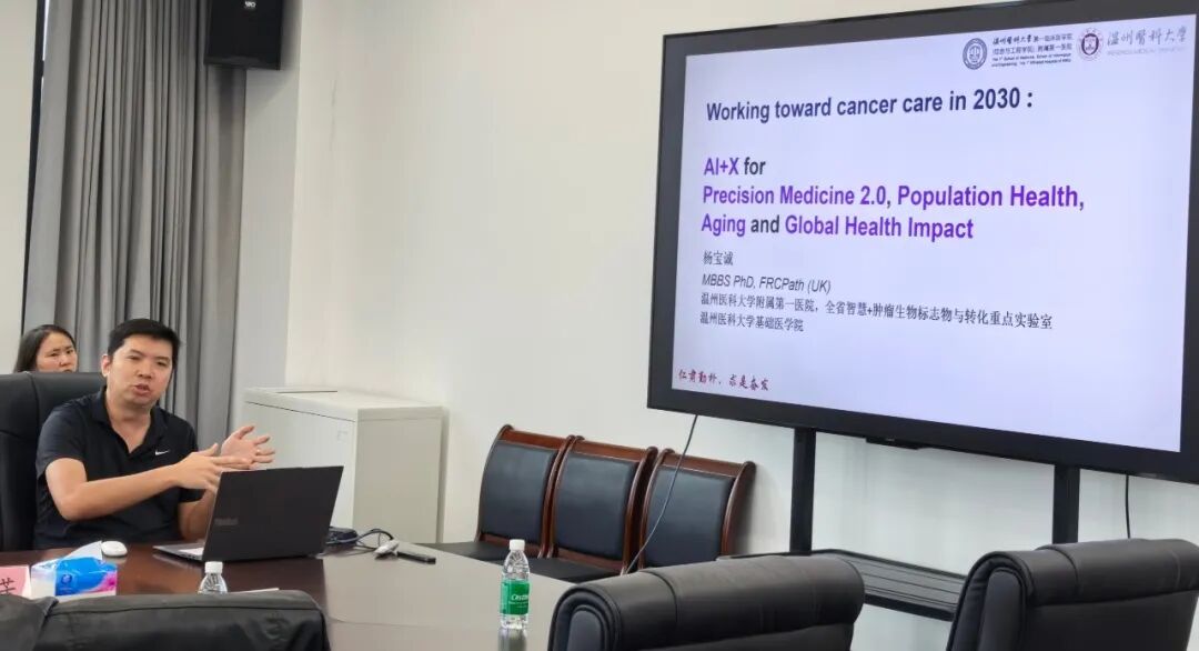
Topic 1: Towards precision diagnosis and treatment of tumors in 2030: AI+X empowers new thinking in tumor pathology in multiple dimensions
Introduction to the speaker
Yang Baocheng, He is a national-level talent from the Ministry of Education of China and a national-level talent from the Ministry of Health of Singapore. He is currently the Director of Immunopathology at Singapore General Hospital, and the project leader and chief scientist of the Singapore A*STAR Institute of Molecular and Cell Biology. A pioneer in the field of international spatial omics, he has published more than 150 academic papers and has been invited to give special lectures at more than 100 international conferences. He concurrently holds core academic positions in many top international tumor immunology societies such as SITC, WIC, ASCO, and ESMO.
Key points of the lecture
This time Based on the forefront of tumor pathology, we propose an "AI+X" multi-dimensional empowerment framework and look forward to the development path of precision tumor diagnosis and treatment in 2030. It was pointed out that traditional pathological diagnosis relies on morphological observation and has a single dimension of information, making it difficult to meet the higher needs of precision medicine in tumor classification, prognosis assessment, and medication guidance. The speaker proposed to use artificial intelligence as the core to integrate multi-source information such as pathological images, molecular markers, imaging features, and clinical data to build an "AI+X" integrated diagnosis model, where "X" represents the expandable dimensions of pathology, molecules, imaging, and clinical. With the help of AI's deep learning and mining of multi-modal big data, pathological diagnosis will move from qualitative to quantitative, single-modal to cross-scale integration, assisted film reading to full-process intelligent decision-making. The lecture describes the vision: By 2030, AI will be deeply integrated into the entire process of material collection, production, preliminary screening, classification, and reporting. Pathologists and intelligent systems will collaborate to make judgments to achieve more accurate, efficient, and personalized tumor diagnosis and treatment, providing a systematic idea for the transformation of pathology departments in the AI era. 。
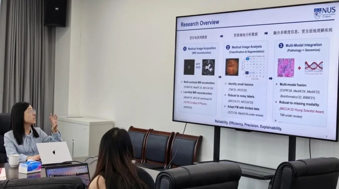
Topic 2: Using artificial intelligence to connect medical imaging and multi-omics to promote precision medicine
Introduction to the speaker
Xing Xiaohan, Dual assistant professor at the Department of Diagnostic Radiology and Department of Electronic Engineering at the National University of Singapore, PhD at the Chinese University of Hong Kong, and postdoctoral fellow at Stanford University in the United States. He has been deeply involved in the field of medical artificial intelligence for a long time, focusing on medical imaging analysis, multi-modal learning and precision medicine, focusing on the fusion modeling of medical imaging, digital pathology, genomics and other multi-source medical data, relying on AI to empower accurate disease diagnosis, prognosis assessment and clinical treatment decision-making. He has published many papers in top international journals and conferences such as Proceedings of the IEEE, IEEE TMI, Medical Image Analysis, CVPR, ICCV, and MICCAI, and won many important academic honors such as MICCAI Young Scientist Award and ASTRO Best Physics Award. 。
Key points of the lecture
This time Focus on the linking role of artificial intelligence in the intersection of medical imaging and multi-omics, and explore the upgrade path of precision medicine. Medical imaging contains rich tumor phenotype information, and multi-omics data such as genome, transcriptome, and proteome reveal tumor molecular mechanisms. However, the two are relatively separated in research and clinical practice, making it difficult to collaborate. Artificial intelligence, especially deep learning, provides a key means to break through this bottleneck. The speaker introduced that AI is used to extract high-dimensional quantitative features of images (radiomics), conduct cross-modal correlation modeling with multi-omics data, and analyze the biological connotations behind image phenotypes. This model can rely on non-invasive or minimally invasive imaging methods to partially replace invasive molecular testing and support non-invasive tumor diagnosis, molecular typing, efficacy prediction and drug resistance monitoring. The lecture also introduced the application scenarios of this technology in personalized diagnosis and treatment, including treatment plan selection, dynamic efficacy evaluation, and recurrence risk warning. It was proposed to build a radiomics-driven precision medical closed loop, using AI to connect imaging and multi-omics to provide patients with more accurate, minimally invasive, and individualized diagnosis and treatment plans. 。
This lecture proceeds step by step, focusing on the cutting-edge direction of precision diagnosis and treatment of tumors. It introduces "AI+X" pathological integrated diagnosis and imaging-multi-omics AI fusion new model respectively, focusing on the core issues of AI empowering tumor diagnosis and clinical decision-making, and broadening research ideas for scientific researchers. Participants responded well and gained a lot 。


Number
023-68059937
Email:Jinfeng@jflab.ac.cn
Address:
No.313, Bella Road Chongqing High-tech Zone
Postal code: 401329

 渝公网安备50009802002274